Introduction
CONCEPT AND MAIN CLASSES
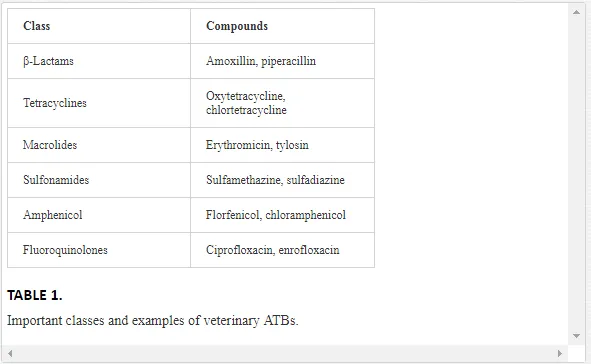
Antibiotics (ATBs) are natural or synthetic chemical agents that belong to the group of drugs that play a major role in the prevention and treatment of diseases in human and veterinary medicine [1] inhibiting (bacteriostatic) or killing (bactericidal) microorganisms such as bacteria, fungi, and protozoa [2], besides acting as animal growth promoters [1]. ATBs differ by their chemical structure and mechanism of action, two characteristics that allow these compounds to be grouped into several classes, such as β-lactams, quinolones, tetracyclines, macrolides, sulfonamides, and chloramphenicol, among others. Some of the main ATBs classes used in veterinary medicine, as well as some examples of compounds belonging to them, are shown in Table 1.
ATBs may be of natural or synthetic origin. The first ATB, penicillin, which is produced by fungi of the Penicillium genus, that is, of natural origin, was discovered in 1929 by the physician and bacteriologist Alexander Fleming. Currently, the ATBs, that are small molecules with molecular weight of less than 1000 Da, are produced by chemical synthesis or by chemical modification of naturally occurring compounds [1].
The use of antibiotics in veterinary medicine, characteristics, and environmental contamination
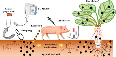
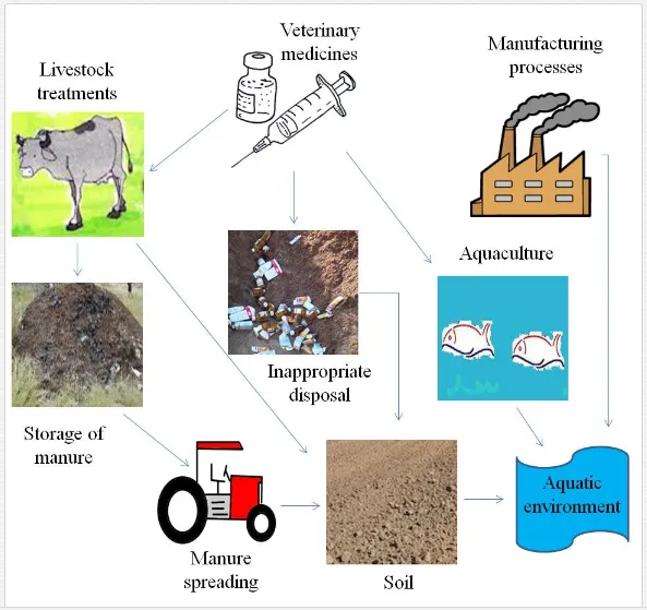
The use of ATBs in the veterinary sector has been for many years an effective method used in animal husbandry, as these chemical agents promote animal growth, besides prevention and therapy against microorganisms [1]. Virtually, livestock activities, such as cattle, pigs, goats, and aquaculture, among others, make the use of these molecules to ensure animals’ good quality and well-being, and in the case of activity for commerce, ensure product quality and market competitiveness. Once ATBs are used and subsequently absorbed by animals, such compounds are metabolized. The metabolism degree depends on the type of substance and the treated species, as well as its age and health condition. If the compound is not metabolized, it will be eliminated in the feces and urine, reaching the environment (mainly soil and water) [3]. According to Kümmerer [1], 80% to 90% of ATBs are excreted as parent compounds in the environment, i.e., compounds that have not undergone metabolism in the animal body. According to Katz [4], the amount of eliminated ATBs varies according to the type of ATB, the dosage, and the type and age of the animal. In the case of farm animals such as cattle, pigs, goats, and sheep, after reaching the soil through feces and urine, this group of drugs may be leached or suffer runoff to aquatic environments and still be in the soil [5]. Three other important soil and water introduction pathways to be mentioned are the packaging disposal in inappropriate places, especially on small farms, where the educational level of most farmers is low, the use of animal excreta for fertilization [6] and water direct contamination through aquaculture. The ATBs may also contaminate the environment by emissions during their fabrication process, although researchers consider this introduction pathway to be less relevant than those described above [7]. Thus, concern about the effects of these compounds in the environment has increased in recent years, which puts them as major environmental concern contaminants. Figure 1 exemplifies the main contamination pathways of veterinary ATBs in the environment.
f all animal husbandry activities, fish farming may be the one that contributes with the largest share of direct contamination of ATBs in the environment. In general, these compounds are administered in fish farming through three forms: inclusion in food (the most practiced and used in tank-nets crops), baths (restricted to water-soluble compounds and administered in tanks with interrupted water renovation during treatment), and finally through hypodermic injection (high cost). According to Shao [8], ATB inclusion in the feed is the most convenient way due to lower amounts of drug required in comparison with administration through water, for example. Consequently, the number of such molecules that enter the aquatic environment would be lower, according to this author.
Cravedi [9] reported that about 7% to 9% of the ingested oxytetracycline was absorbed during the passage through the gastrointestinal tract of rainbow trout, and thus 93% to 99% polluted the environment. Rogstad et al. [10] observed absorption of less than 1% after 24 h of oxytetracycline administration and of 2.6% after 72 h. Among sick fish, low intake rate is common due to reduced palatability of the diet. Thinking of decreasing the contamination of aquatic environments, hypodermic injection or vaccine has been used in many fish farms. Even with the use of ATBs in aquaculture facilities, limited data on types and amount used of these products are not available. In the study of Sapkota et al. [11], a list of 26 antibiotics used in the 15 countries that more practiced aquaculture by the year of 2005 according to FAO was presented, which comprises representatives of the class of sulfonamides, tetracyclines, penicillins, quinolones, nitrofurans, macrolides, aminoglycosides, and chloramphenicols. In general, ATBs used in aquaculture are oxytetracycline, florfenicol sarafloxacin, erythromycin and sulfonamides potentiated with trimethoprim or ormetoprim [12]. In Brazilian fish farms, only florfenicol-based ATBs are approved by the Ministry of Livestock Supply (MAPA) for use in tilapia [13], although others, such as oxytetracycline, are also used. Both are representatives of the classes of chloramphenicols and tetracycline, respectively, and the main form of use of these drugs has been through inclusion in the feed. Florfenicol is a fluorinated analog of thiamphenicol [14], which binds to 50S and 70S ribosome subunits [15], inhibiting protein synthesis transpeptidation of Gram-negative and Gram-positive bacteria [16]. Oxytetracycline is an antibacterial agent, which is effective in the treatment of infections caused by Gram-positive and Gram-negative bacteria, mycoplasmas, and large viruses. It inhibits protein synthesis by preventing the association of aminoacyl-tRNA to the bacterial ribosome [3].
As with most chemical agents, the destination and behavior of ATBs in the environment is influenced by the physical and chemical characteristics of the compounds (molecular structure, size, shape, solubility, hydrophobicity) and of the soil (pH, texture field organic), besides the climatic conditions (temperature, precipitation) [17] and biological factors (microbial degradation). ATBs that have high potential of sorption (Kd) to the soil particles, for example, tend to accumulate and persist in this matrix, unlike those who have low Kd value, which are easily transported to aquatic environments [18]. According to Regitano and Leal [19], in general, compounds with Kd <5 L kg-1 values and half-life of less than 21 days, such as sulfonamides (Kd = 0.2 to 2.0 L kg-1), for example, have relative persistence and can be leached into groundwater, unlike those that have Kd > 5 L kg-1 and half-life of more than 21 days, which tend to accumulate in the soil surface layers, as is the case of compounds belonging to the group of tetracyclines and fluoroquinolones (Kd = 70 at 5.000 L kg-1). Kd values may vary considerably for certain compounds in different types of soil [20]. According to Tolls [21], ATBs sorption may also be influenced by cation exchange processes, by adsorption to the surfaces of clay minerals, by complexing reactions with metal ions and by hydrogen bridges. For example, in the study of Sassman and Lee [22], the main mechanism involved in the sorption of tetracyclines was cation exchange, and sorption potential was influenced by the environment pH and by the cation exchange capacity of clay minerals prevailing in the soil matrix. ATBs, which are mostly complex molecules, may have different functionalities within the same molecule, which causes that under different pH conditions they can be neutral, cationic, anionic, or zwitterionic. Due to different functions within a single molecule, its physicochemical and biological characteristics, such as the log Kow [23], sorption behavior, photo reactivity, antibiotic activity, and toxicity, can change with pH. Other factors that are pH dependent are solubility, hydrophobicity, and log Kow. Regarding drugs solubility being pH dependent, this can affect not only destination and transport, but also the assessment of environmental effects, which includes toxicological assessments [24], which are going to be portrayed in this chapter.
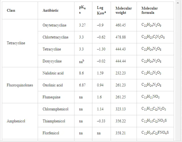
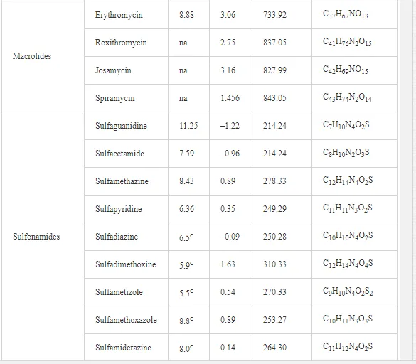
Generally, pharmaceutical products are compounds characterized by a complex chemical structure that have very variable molar masses (172 at 916 g mol-1), low volatilization potential, several ionizable functional groups (amphoteric molecules), different pKa values, and low octanol–water partition coefficient values (log Kow), which indicates low bioaccumulation potential [25]. The log Kow indicates the tendency of an organic chemical product to partition into lipids or fats and adsorb to particles of soils, sediments, biomasses, and muds [23]. Table 2 shows some of these characteristics described above for some ATBs.
Occurrence in the environment
SURFACE WATER, GROUNDWATER, AND SEDIMENT
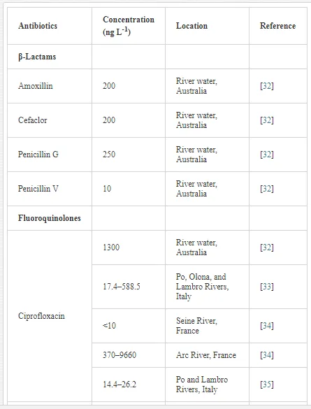
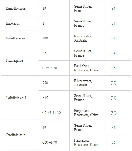
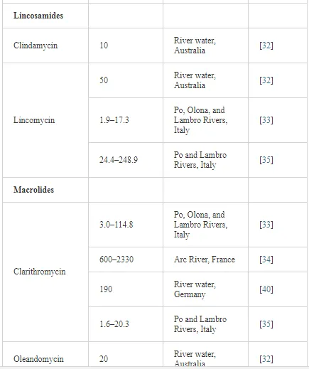
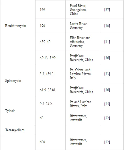
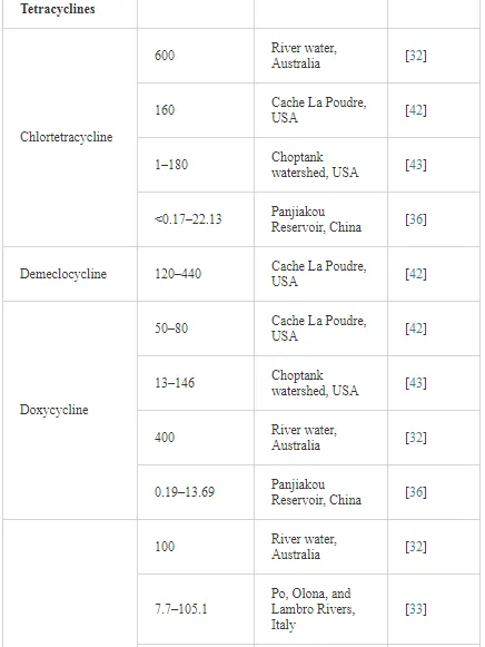
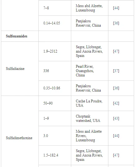
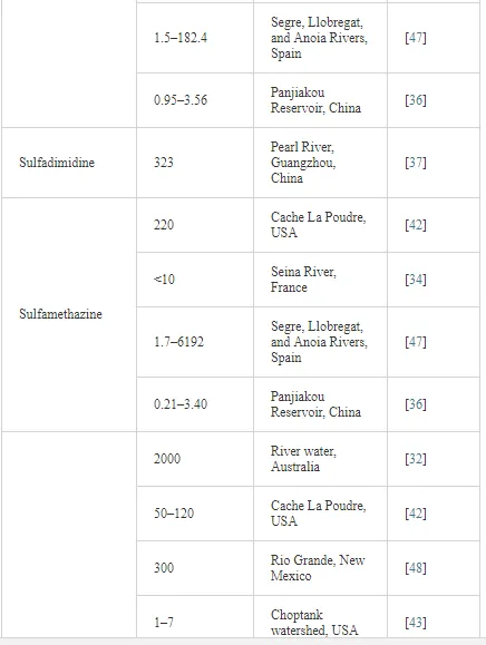
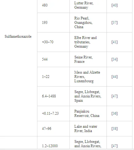
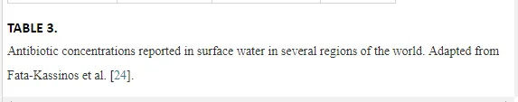
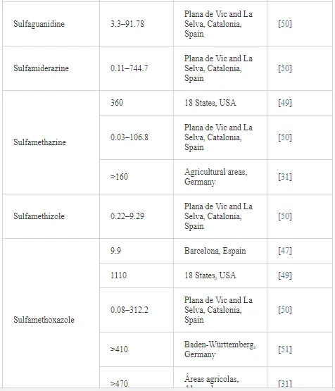
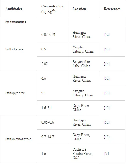
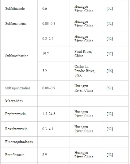
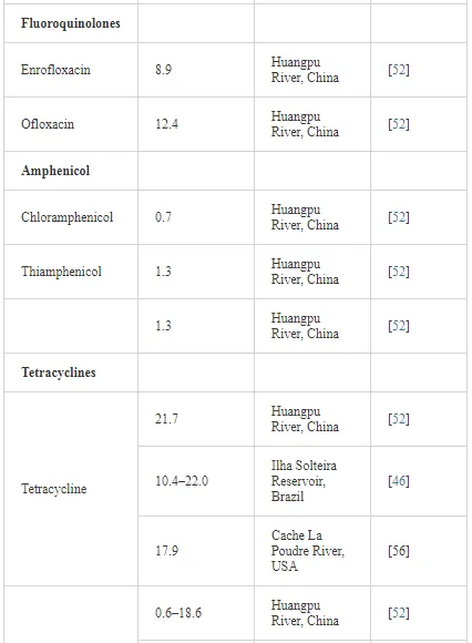
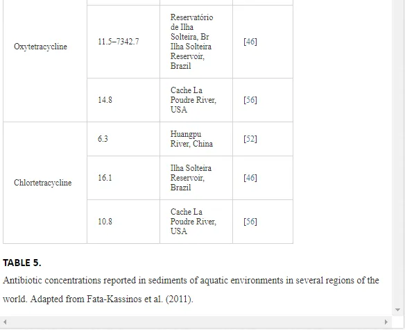
As described previously, veterinary ATBs may contaminate the environment after their use, principally soil and water matrices, and also aquatic nontargeted organisms and sediments. The first reported case of antibiotic contamination in surface waters happened in England more than two decades ago, when Watts et al. [27] found at least one compound belonging to the group of macrolides, tetracyclines, and sulfonamides in river water, in 1 µg L-1 concentrations. Subsequently, other studies, such as those of Richardson and Bowron [28], Pearson and Inglis [29], Ternes [30], and Hirsch et al. [31], have been developed, enabling the detection of other ATBs groups. Although the study of pharmaceutical residues in the environment is relatively a new topic, a lot of papers have already been published from the 1990s to the present day, as can be seen in Tables 3, 4, and 5, which describe ATBs and their reported concentrations in different environmental matrices in several parts of the world.


Antibiotic effects on the environment
Veterinary ATBs are designed to affect microorganisms found in animals. However, as discussed above, they are rapidly eliminated in its active form or as by-products, contaminating the environment. After contaminating the environment, such drugs have the potential to cause adverse effects to the aquatic and terrestrial biota of different trophic levels and also to humans, through consumption of contaminated food derived from aquaculture or through contact with contaminated water. ATBs transference in the body is determined by its ability to move through the lipid bilayer of epithelial cells. The most important properties affecting their permeation across biological membranes are lipophilicity, hydrogen bonding capacity, size, and charge [58]. To demonstrate the negative effects of these compounds, several authors have performed toxicity tests using a wide range of test organisms under controlled conditions [59–70]. Toxicity tests are divided into acute and chronic. The acute toxicity test is designed to evaluate the effects on organisms in a short period of exposure, with the goal of determining the concentration of a studied substance that produces deleterious effects in controlled conditions. When the test organism is fish, lethal effect is observed in most of the times, from which the concentration of the toxic agent that causes 50% mortality (LC50) is determined. On the other hand, for microcrustaceans, the observed effect can be lethality and also mobility, and in the latter case, the average effective concentration (EC50) that causes 50% immobility is calculated [71].
In chronic toxicity tests, organisms are continually exposed to the evaluated substance for a significant period of time of their life cycle, which can range from half to two thirds of the cycle [71]. Depending on the tested substance characteristics, due to the long test period, it may be necessary to the test solutions to be renewed. In this test, sublethal effects, such as changes in growth and reproduction, changes in behavior (such as movement difficulty and increased lid opening frequency, in the latter case to fish), physiological, biochemical, and tissue alterations [72, 73], among others are evaluated. The chronic toxicity test depends directly on the results of the acute test, once sublethal concentrations are calculated from CL50 and CE50. For choosing the test organism, the following selection criteria are often used: abundance and availability, significant ecological representation, cosmopolitanism, knowledge of its biology, physiology and diet, genetic stability and uniformity of their populations, sensitivity, commercial importance, ease of cultivation in the laboratory and, if possible, the species should be native, to a better representation of ecosystems [71]. The sensitivity of algae to ATBs varies widely. In a performed toxicity test, it was shown that the green alga Selenastrum capricornutum was less sensitive than Microcystis aeruginosa microalgae for most of the tested molecules. The growth of M. aeruginosa was inhibited when concentrations of less than 0.1 mg L-1 were exposed [66]. Blue-green algae (cyanobacteria) were also sensitive to several ATBs, such as amoxicillin, penicillin benzyl, spiramycin, tetracycline, among others. All these results are very worrying, once that algae are located at the base of the food chain, and a drop in the population of these organisms can disrupt aquatic ecosystems. Reproductive effects have also been observed in aquatic organisms, such as Artemia sp. and Daphnia magna when exposed to ATBs [55, 61, 66, 74, 75]. It is important to consider that reproductive effects in any population of organisms can cause considerable damage to the natural balance since the organisms are directly related to each other in the trophic chain.
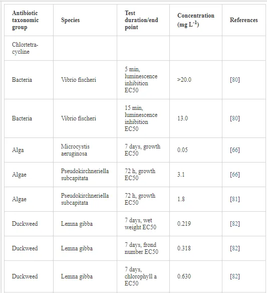
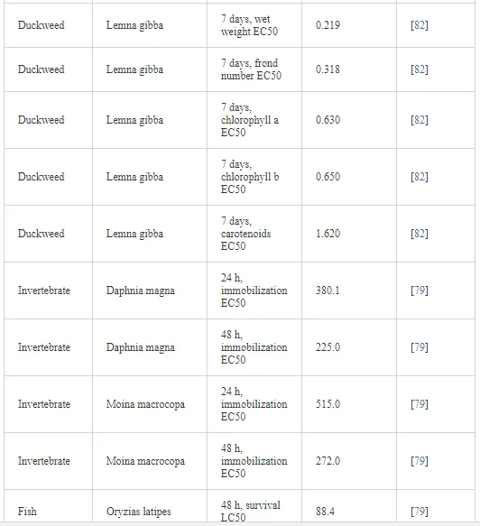
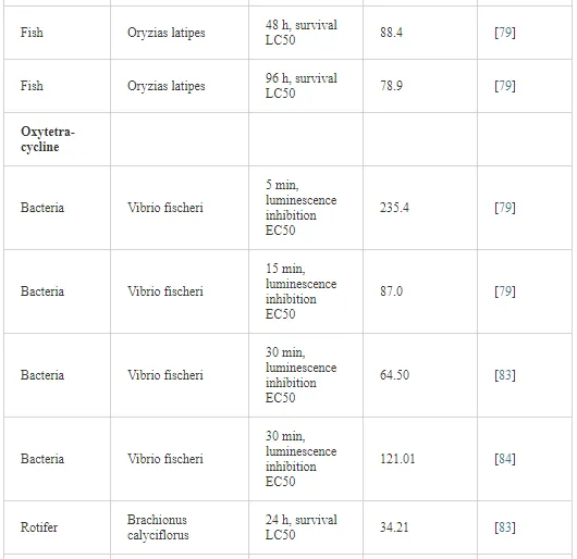
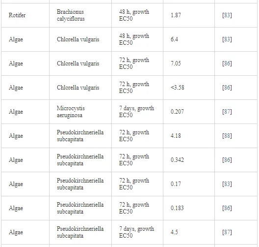
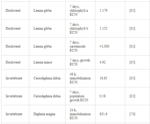
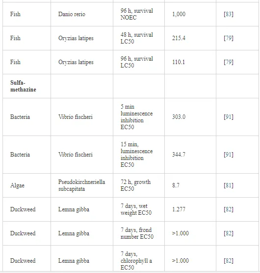
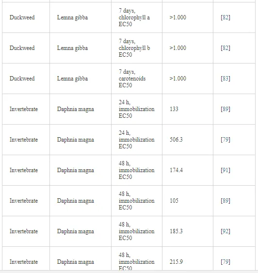
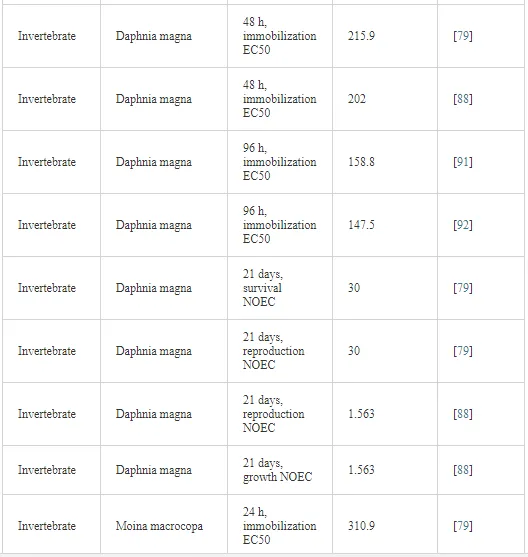
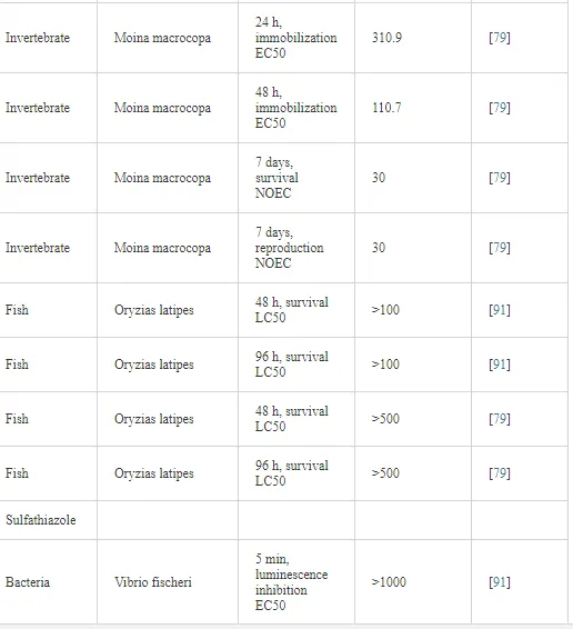
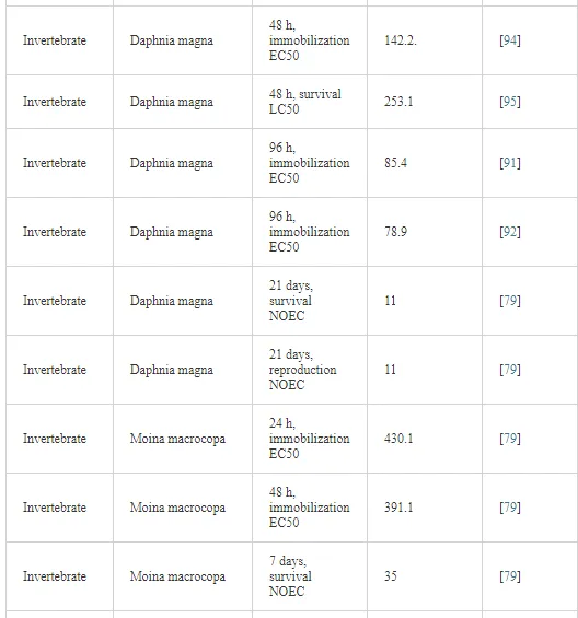
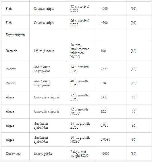
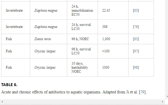
Numerous studies have evaluated the acute toxicity of ATBs for different aquatic organisms. For example, Wollenberger et al. [75] studied the acute toxicity of nine commonly used veterinary ATBs and reported lower acute toxicity (CE5048h, mg L-1) of the oxolinic acid (4.6) and higher toxicity to oxytetracycline (~1000). Previously, Dojmi di Delupis et al. [76] had reported moderate toxicity to aminosidine, bacitracin, erythromycin, and moderate lincomycin ATBs to D. magna microcrustacean, with CE5048h value of between 30 and 500 mg L-1, with bacitracin being the most toxic. In another study, Kolodziejska et al. [77] determined the toxicity of four veterinary ATBs for different aquatic organisms. In this study, oxytetracycline and florfenicol had stronger effects on Lemna minor (CE50 = 3.26 and 2.96 mg L-1, respectively) and on green alga Scenedesmus vacuolatus (CE50 = 40.4 and 18.0 mg L-1) than on the marine bacterium Vibrio fischeri (CE50 = 108 and 29.4 mg L-1) and on microcrustacean D. magna (CE50 = 114 and 337 mg L-1). The chronic effects of ATBs to aquatic organisms were also studied. Kin et al. [78] evaluated the chronic toxicity of acetaminophen and lincomycin ATBs for two crustaceans species (D. magna and Moina macrocopa) and for the fish Oryzias latipes. To D. magna, acetaminophen ATB caused no significant effect on reproduction when exposed to the concentration of 5.72 mg L-1. Similar results were observed for survival and growth when microcrustaceans were exposed to the highest concentration of lincomycin (153 mg L-1). For fish, a significant reduction in survival was observed 30 days after hatching, when exposed to 95 mg L-1 of acetaminophen and 0.42 mg L-1 of lincomycin. Several other studies were conducted to evaluate ATBs acute and chronic toxicity of different classes, using organisms of different trophic levels, as can be seen in Table 6.


Genotoxic and enzymatic effects on aquatic organisms exposed to ATBs were also observed by several authors. For example, Botelho et al. (submitted manuscript) reported genotoxic effects of oxytetracycline and florfenicol ATBs in concentrations found in the water of a major Brazilian reservoir where fish farming activity is practiced with Oreochromis niloticus fish species. In this study, DNA damage was observed using the comet test when exposed to concentrations of 425 and 4000 ng L-1 of florfenicol and oxytetracycline, respectively. Oliveira et al. [98] observed the inhibition of catalase activity in adult brain and gills of Danio rerio fish when exposed to higher concentrations of amoxicillin (50 and 100 mg L-1). There was also a tendency for the induction of glutathione S-transferase (GST) enzyme at all concentrations of the same ATB. In this same study, a dose-dependent catalase was observed in the brain of D. rerio adults after oxytetracycline exposure, while GST activity increased after exposure to concentrations higher than 1 mg L-1 of oxytetracycline in muscle and liver samples.
Most of the studies related to ATBs effects on aquatic organisms refer to acute effects (mainly lethality) in a short period of time. Note that in the aquatic environment, due to the phenomenon of dilution, the concentrations of chemicals in general, including ATBs, are found at the levels of µg L-1 and ng-1. Thus, the observed effects will be chronic, i.e., at a considerably longer period than that observed for acute effects. Thus, in toxicity evaluations, especially to aquatic organisms, the use of environmentally relevant concentrations should be taken into account since this way the effects will be more realistic and will portray in a more real way what happens in the environment if such chemical agents are present. Soil plays important roles in ecosystems since it is the basis of nutrients and the animal and plants habitat, in addition to functioning as an immense bioreactor, where the degradation of pollutants and nutrients transformation occurs. However, as already seen in this chapter, the soil may also be the final destination of ATBs used in veterinary medicine originating from manure and sewage mud used to fertilize vegetables [99] or from package disposal. Due to the ecological importance of soil for the ecosystem, it is important to know whether or not ATBs have negative effects on the fauna.
As shown earlier in this chapter, once in the soil, depending on the physical and chemical characteristics of the ATBs and the soil, they may follow different pathways, such as being leached or carried superficially by rain, contaminating aquatic environments (low Kd values) or persisting in the soil (high Kd values).
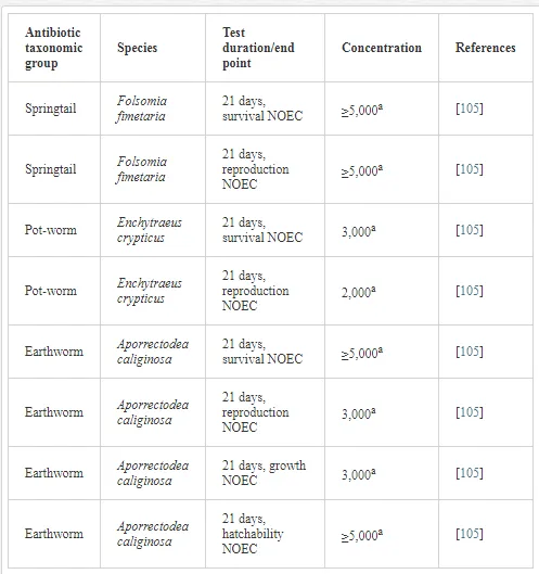
In general, the effects of ATBs to aquatic organisms are higher than those of soil fauna, and thus little is known about the toxicity of these drugs for these organisms. According to Ding and He [100], once in the soil, ATBs can change the structure of the microbial community because even to those which have a broad spectrum of action, selective effects on several microorganism groups may occur. As a result, the relative abundance of microorganisms is changed, interfering with the interactions between different species. The sorption of pollutants in general in the soil is one of the major mechanisms controlling toxicity, by reducing its availability [101]. Thus, in toxicity studies with chemical agents, the choice of a molecule with low Kd is recommended. In addition, toxicity to organisms in the soil decreases over time due to transformations the molecule undergoes over time through less toxic secondary compounds and due to tolerance of some soil microorganisms to ATBs [102], such as some Pseudomonas species, for example [103]. Girard et al. [104] studied the effects of ciprofloxacin ATB on soil microbial communities and observed a reduction in soil microbial activity in the first 25 days of experiments, when exposed to concentrations ranging from 0.2 to 20 mg kg-1. According to the authors, this behavior is due to this molecule being bacteriostatic. From this result, according to the authors, ciprofloxacin could interfere with the recycling of nutrients in the soil. In Table 7, some studies that were conducted with oxytetracycline ATB for three organisms that live in the soil can be observed
Generally, veterinary ATBs can suffer abiotic or biotic degradation on soil–water compartment. However, some degradation products have similar toxicity to the parent compound [106]. Degradation can be affected by environmental conditions, such as temperature, humidity, season, soil type, pH, and characteristics of the molecule, such as size, among others. With respect to the season, for example, in winter, the degradation half-life of ivermectin is six times higher than in summer, and degradation was faster in sandy soil than in sandy loam soil [107, 108].
Microbial resistance
One of the biggest problems related to the use of ATBs, in addition to those already discussed in this chapter, is the development of bacterial strains resistant to ATBs in the environment, mainly due to the continuing use of these drugs at low concentrations. Bacterial resistance arises and is maintained by mutations in the bacterial DNA or by horizontal gene transfer mechanisms, which include conjugation with other bacteria, transduction with the bacteriophage, and free DNA uptake via transformation.59 In the case of continuous and prolonged use of sublethal concentrations and the subsequent elimination of feces in the soil, they could cause the sharing of resistant plasmids to nonresistant organisms [18, 99]. Another possibility of occurrence of bacterial resistance is that low concentrations of ATB residues transferred to the soil by the application of contaminated animal manure favor the selection of resistant populations [109]. However, the direct introduction of resistant microorganisms derived from feces of animals treated with ATBs seems to be more important to resistance [99] than induction due to the presence of ATB residues on the environment. It is important to remember that there is a large reservoir of ATBs-resistant bacterial genes in the soil. However, according to Schmidt e al. [110], it is not known whether this occurs naturally or due to the use of veterinary ATBs. As an example, in a study by Esiobu et al. [102], isolated bacteria of a garden soil fertilized with dairy cattle manure showed 70% resistance to ampicillin, penicillin, tetracycline, vancomycin, and streptomycin ATBs.The exposure intensity of bacteria to ATBs agents influences the amplitude of its resistance, and the exposure intensity usually depends on the origin of the treatments by which bacteria were submitted. Costanzo et al. [111] indicated that bacteria from a sewage treatment plant reactor were resistant to ciprofloxacin, tetracycline, ampicillin, trimethoprim, erythromycin, and sulfamethoxazole antibiotics, while bacteria isolated from the effluent receiver watercourse showed resistance to erythromycin and ampicillin. This same study showed that erythromycin, clarithromycin, and amoxicillin ATBs, at a concentration of 1.000 μg L-1, decreased more significantly the rate of bacterial denitrifying.
In aquaculture, the intensive use of ATBs provides a selective pressure for the creation of bacteria resistant to drugs and genes resistant to transmitted pathogens of fish and other bacteria in the aquatic environment. From these resistant bacteria, resistance genes can be spread by horizontal gene transfer and transfer to human pathogens. Drug-resistant pathogens present in the aquatic environment can directly reach humans. The horizontal gene transfer can occur in the aquaculture environment, in the food chain, or in the human intestinal tract. Among ATBs commonly used in aquaculture, several are classified by the World Health Organization (WHO) as extremely important for use in humans. The occurrence of ATBs resistance in human pathogens severely limits the therapeutic options in human infections. Taking into account the rapid growth and the importance of the aquaculture sector in many regions of the world, due to the widespread, intense and often irregular use of ATBs in this animal production area, efforts are necessary to prevent the development and spread of bacterial resistance in order to reduce the risk to human health [112]. Another issue in aquaculture regarding bacterial resistance needs to be highlighted, that is, if bacterial populations are resistant to a certain ATB used in this sector, or the producer changes the ATB or increases the dose in anticipation of a more efficient control. However, these two practices make such microorganisms to become resistant to this new applied molecule over time.
Increasing the dose may also have negative effects on native aquatic biota of where the creation is installed. In the study of Akinbowale et al. [113], isolated bacteria from water samples and organisms used in aquaculture showed widespread resistance to ampicillin, amoxicillin, cephalexin, and erythromycin ATBs; frequent resistance to oxytetracycline, tetracycline, nalidixic acid, and representatives of the sulfonamide group; and infrequent resistance to florfenicol, chloramphenicol, ceftiofur, oxolinic acid, gentamicin, and trimethoprim. In another study performed on the Ilha Solteira Reservoir, São Paulo, where one of the largest and most important aquaculture parks in Brazil is located and where fish farming is intense, Monteiro [46] studied the bacterial resistance in Nile tilapia kidneys, which is a species cultivated in this place, and observed bacterial resistance to sulfonamides, quinolones, and tetracyclines. These two examples from cited studies confirm that both aquaculture products and water from aquaculture environments have risks of transferring ATBs-resistant bacteria to humans through these product consumption and contact with water, as mentioned in the previous paragraph. Remember that in the case of the Ilha Solteira Reservoir, this environment is an important aquaculture redoubt where the population uses its waters for water sports, in addition to fishing and fish consumption. As a direct consequence of bacterial resistance, there is the increased frequency of ineffective treatments, increased severity of infections, prolonged duration of diseases, increased frequency of bloodstream infection, increased hospitalization, and increased mortality. The prolongation of diseases has been demonstrated in case-control studies of Campylobacter resistant to fluoroquinolones, and the increased severity of infections of Salmonella typhimurium resistant to quinolones was also demonstrated, as well as increased morbidity or mortality also assigned to nontyphoidal Salmonellaserotypes and to Campylobacter [112].
Several studies have reported the occurrence of bacterial resistance in environmental compartments, such as in wastewater, groundwater, surface water, sediments, and soils [114–119]. As discussed so far, bacterial resistance is a threat to the effectiveness of ATBs in animal husbandry and to the health of the environment. Therefore, the prudent use of these molecules in all livestock sectors seems to be the solution to combat or reduce this problem.
Detection and quantification of antibiotics into the environment
SAMPLE PREPARATION TECHNIQUES
The determination of antibiotics in environmental samples is a difficult task due to the high complexity of the analyzed matrices and the low concentrations of these compounds in the samples [120].The sample preparation step affects all the other steps of the test and therefore is critical for unambiguous identification, confirmation, and quantification of antibiotics. It includes the isolation and/or preconcentration of interest compounds from the matrix and also properly provides the compounds for the separation and detection. Sample preparation takes typically more than 70% of total analysis time. Chromatographic methods are usually preferred in the analysis of organic molecules, which causes the need to have an initial sample preparation, a process of extraction, which is normally a liquid–liquid extraction, followed by a clean-up process, which is usually a solid-phase extraction (SPE). Comparing the analysis by ultrafast chromatography with the conventional, sample pretreatment processes are more laborious and time consuming, as it requires an even purer extract. For this reason, many new sample preparation techniques have been developed, and there is a continuing interest in this area. A quick search in the scientific literature showed that more than 1300 articles on analysis of antibiotic residues were published during the period of 2004–2015, and liquid extraction (LE) and liquid–solid extraction (LSE) were the most popular sample treatment techniques, which were used in 30% and 60% of the reported studies, respectively. The LE includes all techniques based on liquids, such as liquid–liquid extraction (LLE), liquid–liquid microextraction, and pressurized liquid extraction (PLE). LSE includes solid-phase extraction (SPE) and all other procedures based on extraction absorbers, such as solid-phase microextraction (SPME), stir-bar sorptive extraction (SBSE), restricted access material (RAM), turbulent flow chromatography (TFC), dispersive solid-phase extraction (DSPE), and matrix solid-phase dispersion (MSPD). Other techniques for some specific applications are the microwave-assisted extraction (MAE), the ultrasound-assisted extraction (UAE), extraction based on immune affinity, and the technique that use molecular imprinted polymers (MIP).
There have been many changes in sample preparation with the advent of mass spectrometry. Previously, methods of analysis were able to analyze residues of only a limited number of compounds (usually a single class of drugs); but with mass spectrometry, now there is the possibility of residue analysis of many compounds in a single analysis. Although mass spectrometry allows the use of simple and generic cleaning methods, the effective removal of matrix constituents is necessary since these may affect the performance of the mass spectrometer (MS), in particular, by ion suppression. There was also the migration of manual sample preparation to faster techniques with automated processes. The automated preparation of samples can be made online (with sample preparation directly connected to the chromatographic system) or offline (sample preparation is automated, but the sample has to be manually transferred to the chromatographic system). Most analytical methods developed for the antibiotic determination in water use offline solid-phase extraction (SPE) and LC-MS/MS [121–127]. Some studies, however, point toward developing methods with the SPE-LC-MS/MS system, which allows reduction in the sample amount, lower preparation time, and consequently, increase in productivity, in addition to less sample manipulation, decreasing contamination chances [128–132]. The SPE-online system has also been used for the determination of pesticides, hormones, explosives, pharmaceuticals, and personal care products [133–137].
Among the advantages of SPE-online method, it is possible to highlight the small sample volume requirements, making it easier to transport and store. As in most cases, the sampling sites are too distant from laboratories. Several sample preparation steps, such as evaporation and reconstitution, are eliminated, and there is less need for sample handling and processing, which lead to a reduction in analysis time and analyst’s interference, minimizing errors, losses, and sample contamination, which is reflected in better method accuracy and precision values, in addition to a significant reduction in the consumption of organic solvent, contributing to the “green chemistry” [138, 139]. Besides these, the automated preparation of samples has the advantage of performing the clean-up, concentration, and separation of the compound in a closed system. This reduces the sample preparation time, and the whole sample becomes available for analysis, leading to a reduction in detection limits. It also decreases the analyst procedural errors, thereby improving accuracy and reproducibility. Moreover, in automated sample preparation, the cost is also reduced, using less solvent and less personnel. Other advantages include reduced risk of sample contamination and elimination of analyte disposal losses by evaporation or degradation during sample preconcentration. Automatic methods also have some disadvantages, such as increase in initial capital expenditure and risk of increased service downtime due to equipment breakdowns, which require parallel processes to be made in order to reduce the laboratory inactivity.
For the determination of antibiotics in environmental, soil, sediment, and manure solid matrices, among others, different procedures are performed, which involve several techniques, such as accelerated solvent extraction (ASE), pressurized liquid extraction (PLE), ultrasound-assisted extraction (UAE), and microwave-assisted extraction (MAE). The solvent choosing is critical to ensure selectivity and minimize the extraction of other matrix constituents. Better diffusion of the solvent in the matrix interstices by mixing the sample and quartz sand is essential for best performance. The correct use of pH also increases extraction efficiency, and pH acids are generally more indicated because they favor electrostatic repulsion between antibiotics and sediment surface, which are both protonated [140].
Prior to antibiotic chromatographic determination, postextraction sample purification is often necessary to remove interfering (e.g., coextracted organic matter or organic solvent) and to achieve lower quantification limits. The adoption of this strategy leads to substantial improvement in method selectivity, where the cleaning is carried out in most cases by solid-phase extraction (SPE). Used adsorbents differ in composition, chemical properties, and affinity with the analyte [140]. Currently, EAU followed by filtration or centrifugation is the most common procedure. Yang et al. [57] developed a method with EAU using a mixture of acetonitrile and citrate buffer (50:50 v/v) placed in an ultrasound bath for 15 min, repeating this process three times. The extract was then purified by using SPE cartridges in series, SAX (6 mL, 500 mg), and HLB (6 mL, 200 mg); the procedure obtained good recoveries for 14 antibiotics studied in the sediment of Pearl rivers in Guangdong province, China, and was also used by Zhou et al. [141], which increased the number of analyzed antibiotics. Antibiotic extraction from sediment and the transfer of these to an aqueous solution can provide the use of the SPE-online system, increasing the sensitivity of the method, facilitating the preparation of samples, and reducing purification steps, as shown by Monteiro [46] in his PhD study. In this study, the procedure proposed by Yang et al. [57] was used, but the purification step using SPE with HLB cartridges was replaced by SPE-online system, in which a semipreparative precolumn with C8 adsorbent was used, obtaining optimal results for antibiotics of the class of tetracyclines, fluoroquinolones, sulfonamides, and phenicols in sediments collected from tank-nets fish farms.
As for biological samples, such as fish and aquatic plants, for example, one of the main problems for quantitative analysis of pharmaceuticals is that the analyte is typically bound to proteins and peptides, with the consequent need for cleavage of these structures before analysis. Enzymatic digestion is widely accepted as a sample preparation method for analyzing compounds in biological matrices. However, these methods are labor intensive and significantly prolong the examination time [142]. Most methodologies use extraction procedures based on liquid–liquid extraction, with relatively polar solvents and subsequent extraction purification using solid-phase cartridges [143, 144]. Another technique that has been highlighted for the extraction and purification of antibiotics in biological matrices is QuEChERS (quick, easy, cheap, effective, rugged, and safe), which was initially developed by Anastassiades et al. [145] for the determination of pesticide residues in food. It has been adapted and has been used for the determination of other compounds, including antibiotics in fish [54, 146]. The extraction procedure should be appropriate to the intended analysis and the reality of laboratory, so factors such as reagents consumption, availability of skilled labor force, and equipment acquisition are crucial. Simple and rapid methods stand out in this context because they are less dependent on high investments [147, 148].
CONFIRMATORY AND QUANTITATIVE METHODS
For the determination of medicaments, different analytical methods are reported in literature, which are primarily valid for biological matrices, such as blood and tissue [148–150], and some modifications in these methods may be sufficient to environmental samples. However, residual drug analysis in WWTP effluents, rivers, subsoils, sediments, soil, and sludge waters still require the development of more sensitive methods for the detection of concentrations in µg L-1 and ng L-1 range. Separations in environmental chemistry generally involve the two most recognized chromatographic techniques: high-performance liquid chromatography (HPLC) and gas chromatography (GC). Knowledge of physical and chemical properties of the analytes is of utmost importance to avoid problems in quantification, which can be related to side reactions, impurities, or degradation in their structure during the analytical method application. For example, tetracycline antibiotics may irreversibly interact with residual metal ions present in sorbents of solid-phase extraction cartridges based on modified silica with alkyl groups (C18, C8, etc.), and certain metals can catalyze the ring opening of β-lactams. This problem can be solved by adding a chelating agent to the matrix to be extracted (Na2EDTA, for example) or by replacing the cartridges sorbent by polymeric material [57]. The solubility of the analytes in the environmental sample and in the eluting solvent as well as in the mobile phase to be used also deserves attention. For example, some antibiotics form water-insoluble lipophilic complexes in the presence of alkali metal cations. Furthermore, penicillin patterns undergo methanolysis when solubilized in methanol, and should be prepared in acetonitrile or another compatible solvent [57]. During the detection step, when using a mass spectrometer (MS), there may be some sort of fragmentation that is characteristic of the analyte, depending on its pH range. For example, the erythromycin in acidic solution has a mass loss of 18 Da, which corresponds to the loss of a water molecule [57]. One of the critical parameters to be observed during antibiotic determination is referred to the sample pH, because in many cases, the medium pH determines the chemical form of the analyte in solution and thus interferes in the extraction efficiency. For example, the low recovery percentage of quinolones extraction process was improved after acidifying the solution in 2.5 pH. However, it is important to choose a pH range in which degradation of the analytes will not occur [57].
Furthermore, the pH of the mobile phase needs adjustment, in which its value depends on the pKa of the compounds to be analyzed. The recommended buffer concentration is in the range between 2 and 20 mmol L-1 to avoid solubility problems in the mobile phase and to facilitate the ionization mode when using the MS detector [128]. The most commonly used stationary phases in HPLC for separation of organic compounds are of reverse phase (RP) type, which are silica based with C18 groups. Stationary phases with C8 groups may be used for β-blockers and antibiotics (tetracyclines, penicillins, sulfonamides, and macrolides) [128], [57]. The mobile phases used in the RP-HPLC are mixtures of methanol–water (MeOH:H2O) or acetonitrile–water (ACN:H2O) with adjustments of the chromatographic strength and mobile phase selectivity to the obtainment of enough resolution to occur the separation of all chromatographic peaks in minimum analysis time. The addition of modifiers, such as formic acid, ammonium acetate, ammonia, etc., is performed in order to favor the process of analytes ionization by medium pH adjustment, improving their interactions with the mobile phase and the stationary phase. Medium pH control may also be performed by using buffered mobile phases. When the mass spectrometer detector (MS) and the electrospray ionization process (ESI) are used, modifiers may also be used in order to favor the process of analytesionization.
DETECTORS FOR HPLC
Spectrophotometric absorption detectors in the ultraviolet range (UV) and by fluorescence were initially used in HPLC equipment for the analysis of compounds, which are absorbed in the ultraviolet region or are fluorescent. The UV detection was used for the determination of antibiotics from fluoroquinolones class in environmental matrices (hospital effluent) [149], and the fluorescence detection was used for the determination of antibiotics in water, sediment, and fish farm plants samples since they are lower cost equipment comparing to HPLC-MS. However, the limits of detection values obtained for UV detectors are much higher, in the range of µg L-1 to ng L-1, in comparison to the MS detector, that can achieve detection limits in the order of ng L-1 to pg L-1 when used in series (MS/MS), offering also the possibility of confirmation of the analyzed compounds. For environmental analyzes for the purpose of screening, with detection limits in the concentration range of µg L-1, the UV detector can be optimally utilized, besides being used when the concentration of the analyte in the matrix is high, as is the case of the publications cited above [149, 151]. The wide use of HPLC-MS/MS in environmental chemistry is due to the fact that most USEPA official methods use this separation and detection mode [140] due to good limits of detection and the possibility of structural confirmation of the analyzed compounds, besides the robustness of the method. The electrospray ionization process is the most used in the detection by mass spectrometry (MS) because it is a more versatile ionization form that works for analytes with median polarity to very polar and poorly volatile, as is the case for most drugs, or thermally labile analytes, such as certain antibiotics, when compared with the atmospheric pressure chemical ionization (APCI), which uses heating in the range of 300°C–400°C for analytes thermal desorption [57]. Several recent studies [152–155] used analytical methods based on HPLC separation with mass spectrometry detection in series with ESI ionization (HPLC-ESI-MS/MS) for the determination of antibiotics in aqueous matrices. This can be explained by the versatility of this ionization mode, which can be used for analytes with polarities ranging between medium and high, with better detectability by ESI.
The most used mass analyzers for analytes detection are the triple quadrupole (QqQ) for sequential mode (mass in series), the time of flight (TOF), and the ion trap. The TOF type analyzer was used in the determination of drugs (analgesics, antibiotics, β-blockers, and antiepileptics) in surface water, groundwater, and wastewater samples due to having higher detectability, linear dynamic range, and mass accuracy than triple quadrupole type analyzers (QqQ), although the best detection limits were found for the QqQ type analyzer [128]. Recently, the quadrupole time-of-flight hybrid analyzer type (Q-TOF) has been used for providing better resolution and detectability than the conventional quadrupole, thus being applied for identification and quantification of drugs unknown metabolites [57, 128, 139]. In recent years, capillary electrophoresis (CE) has become a popular technique because of its simplicity, high separation power, short analysis time, and low consumption of sample and solvents [138, 139]. Among different CE modes, micellar electrokinetic chromatography (MEKC) [140], which makes use of micellar solutions of ionic surfactants, has proved to be a very attractive technique for separating different medicament classes, including antibiotics, nonsteroidal and steroidal anti-inflammatory agents, and analgesics [141]. However, capillary electrophoresis with UV-Visible detection has not been applied for drug residue analysis in small parts levels per billion (µg L-1) due to its lower intrinsic detectability [46].
TRENDS IN LIQUID CHROMATOGRAPHY APPLIED TO THE STUDY OF ANTIBIOTICS
The trends in high-performance liquid chromatography applied to the study of antibiotics include the use of ultra-performance liquid chromatography (UHPLC), two-dimensional liquid chromatography (LC-LC), and hydrophilic interaction chromatography (HILIC). The UHPLC is one of the latest advances in liquid chromatography, using stationary phases with smaller particle diameters (approximately 2μm) compared to those employed in HPLC. The use of these particles, in addition to high linear velocities of the mobile phase (MP), allows to reduce the analysis time while maintaining resolution and separation efficiency, providing less broad peaks (5–10 s), besides drastically reducing the analysis time to approximately 10 min or less [136].
An example of UHPLC applied to the study of antibiotics is the work done by Zhou et al. [156]. A robust and sensitive method with UHPLC-MS/MS was developed for the simultaneous determination of multiclass of antibiotic residues in several environmental matrices (surface water, pond wastewater, effluent, sediment, sludge, and manure). The analytical method applied SPE with HLB cartridges for water samples and ultrasound extraction for solid samples followed by cleanup using SAX-HLB cartridges. The method was successfully applied to the analysis of environmental samples collected from a WWTP of a swine farm. The detection of several antibiotics with high concentrations in the analyzed samples indicates that WWTP and animal farms are two major sources of antibiotic residues in the environment.
Two-dimensional liquid chromatography is a good alternative when performing analysis in complex samples, such as biological samples, for example, because with the increase of a dimension, there is also the increase of peak capacity, and subsequently the separation process is enhanced [157, 158]. A very interesting example of this technique was the method developed by Wang et al. [159], which analyzed 14 antibiotics in urine with two-dimensional liquid chromatography coupled with Q-TOF mass spectrometer detector, quadrupole time-of-flight hybrid analyzer, where detection limits of 0.04 to 1.99 ng mL-1 were obtained. The method was used to identify the antibiotics in urine of children, but it can be a great tool for use in environmental matrices, especially biological matrices such as fish, crustaceans, aquatic plants, and more.
The term HILIC was proposed by Alpert in 1990 as an acronym for “hydrophilic-interaction chromatography” for the separation of polar solutes. This technique has also been also called “hydrophilic-interaction liquid chromatography,” and “aqueous normal phase.” In a simple way, it can be said that HILIC is an HPLC form very similar to liquid chromatography “in normal phase,” using a column in hydrophilic stationary phase (“normal”), but with an eluent comprising water, buffer, and a high concentration of organic solvent that is miscible with water (typical of a “reverse phase”). The elution order obtained in an HILIC system will be practically opposite to that obtained when employing the reversed phase (RP) mode. Retention is directly proportional to the polarity of the solute and inversely proportional to the polarity of the mobile phase [160]. The HILIC was applied to extract and quantify spectinomycin and lincomycin in manure supernatant liquid from swine and farmland erosion water treated with manure [161]. It was also successfully applied in the determination of oxytetracycline in environmental waters [162], and the results showed that there was no irreversible adsorption of oxytetracycline in the stationary phase, which shows that the silica column may be used in the determination of tetracycline antibiotics in HILIC mode with no peaks distortion, providing a different understanding of what was previously found in the literature. As prospects for the area of liquid chromatography applied to the study of antibiotics, the identification of metabolites and processing products as well as even lower detection ranges and quantification limits is highlighted. HPLC is also closely linked to applied environmental legislation because today, for the vast majority of antibiotics, there is no maximum permissible concentration established by the legislation. Studies on bioremediation and advanced processes of wastewater treatment, such as chemical and photochemical oxidation through advanced oxidation processes (AOP), ozonolysis, nanofiltration, reverse osmosis, membrane bioreactors, etc., are likely to use high-performance liquid chromatography as key tool in monitoring antibiotic degradation processes in artificial or natural environments.
Final remarks
Veterinary antibiotics have become an integral component in maintaining animal health, and although they have been used in large quantities for some decades, the existence of these substances in the environment has received little importance until recently. It is only in recent years that a more complex investigation of antibiotic substances has been undertaken in order to permit an assessment of the environmental risks they may pose. This chapter showed that the fate of antibiotics in soil–water systems and their effects on plants, soil, and aquatic organisms have been heavily studied through chromatographic analysis and toxicity test with many species. The multianalyte methods using SPE, LC/MS, LC/MS/MS, ASE, and others described in the chapter have been used to show the occurrence and transport of antibiotics from their sources into the environment. The environmental dissemination of antibiotic-resistant bacteria and their relationship with human health has also been the objective of this study and needs to be investigated in greater depths by health and regulatory bodies so that a compromise can be made when it comes to the prudent use of VAs and their risk to human health and the environment in general.